Waterproof Medical Endoscope – Ojos Medical's Underwater Capability
In actual clinical scenarios such as perfusion, the underwater environment can easily cause refraction-induced color shift, overexposure from reflected light, and water ingress into the circuit. Overcoming these challenges relies not only on the sealing process of the front‑end module, but also on the precise tuning of the backend image processing board.
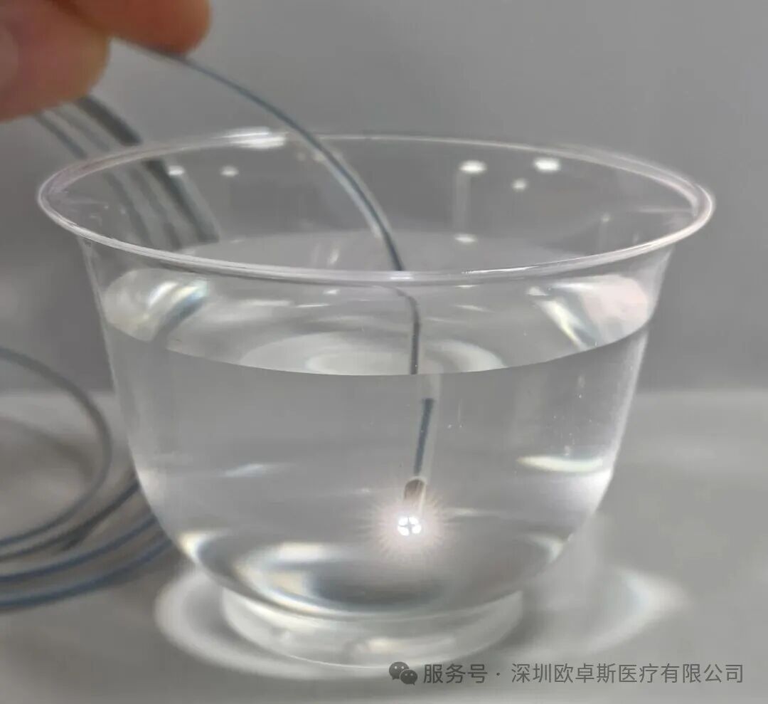
In our previous post, we demonstrated the basic functions and physical structure of the module and image board in normal conditions. Today, we are taking it a step further – fully submerging the module in water to test its sealing performance and the three core functions of the image board in a realistic underwater environment.
Underwater Sealing & Illumination Test
The module and its cable were fully immersed in a water cup and powered on. The module lit up successfully underwater, with the front‑end LED illumination working properly – no flickering, short circuit, or fogging observed. This demonstrates excellent precision sealing and waterproof reliability.
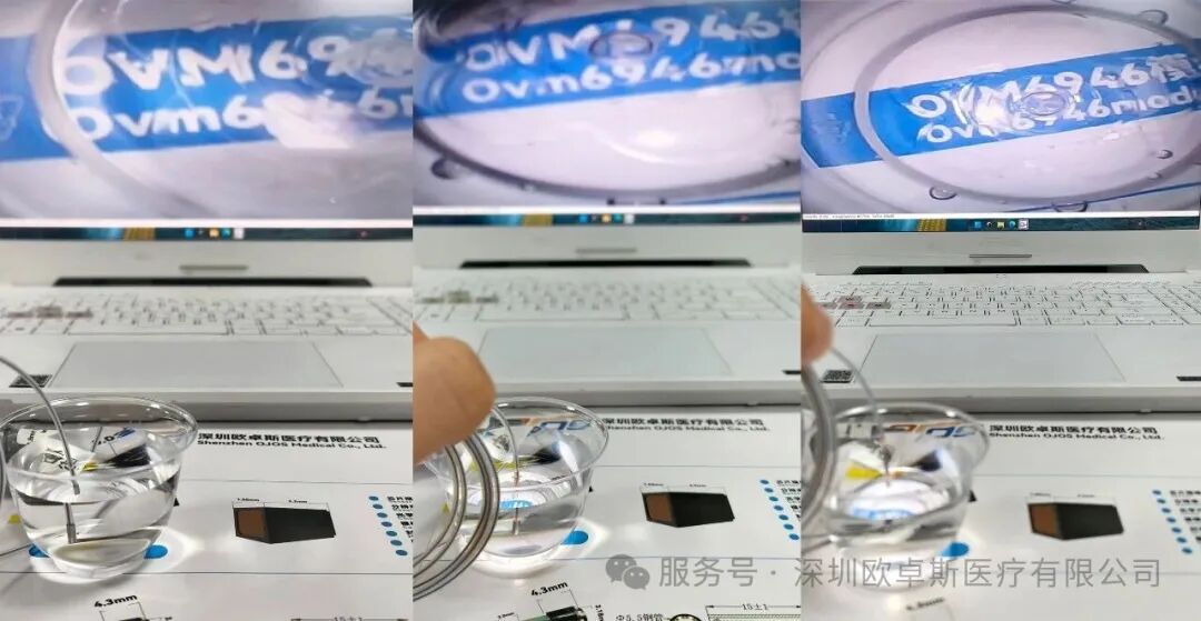
Using the dimming controls on the image board, we tested the module's brightness in steps – from low‑light fill to full‑power illumination. The transition was smooth and stepless, with no flickering, showing outstanding light intensity control underwater and the flexibility to adapt to various lighting conditions.
White Balance & Color Accuracy Test
Underwater refraction and lighting can easily cause color shifts. By adjusting the white balance function on the image board, color deviations can be effectively corrected. On the test screen, the text and icons behind the module were clearly displayed with accurate, natural colors – clean and undistorted.
Real‑Time Image Capture
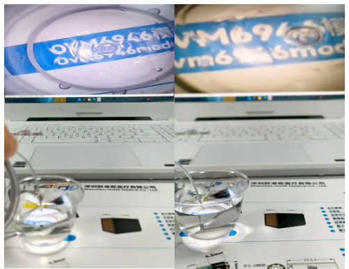
During dynamic underwater observation, pressing the capture button on the image board instantly froze the current high‑definition image. The on‑screen pop‑up responded quickly, with a clear, motion‑blur‑free freeze‑frame – making it convenient for operators to save critical image details during procedures.

